What we do
Research
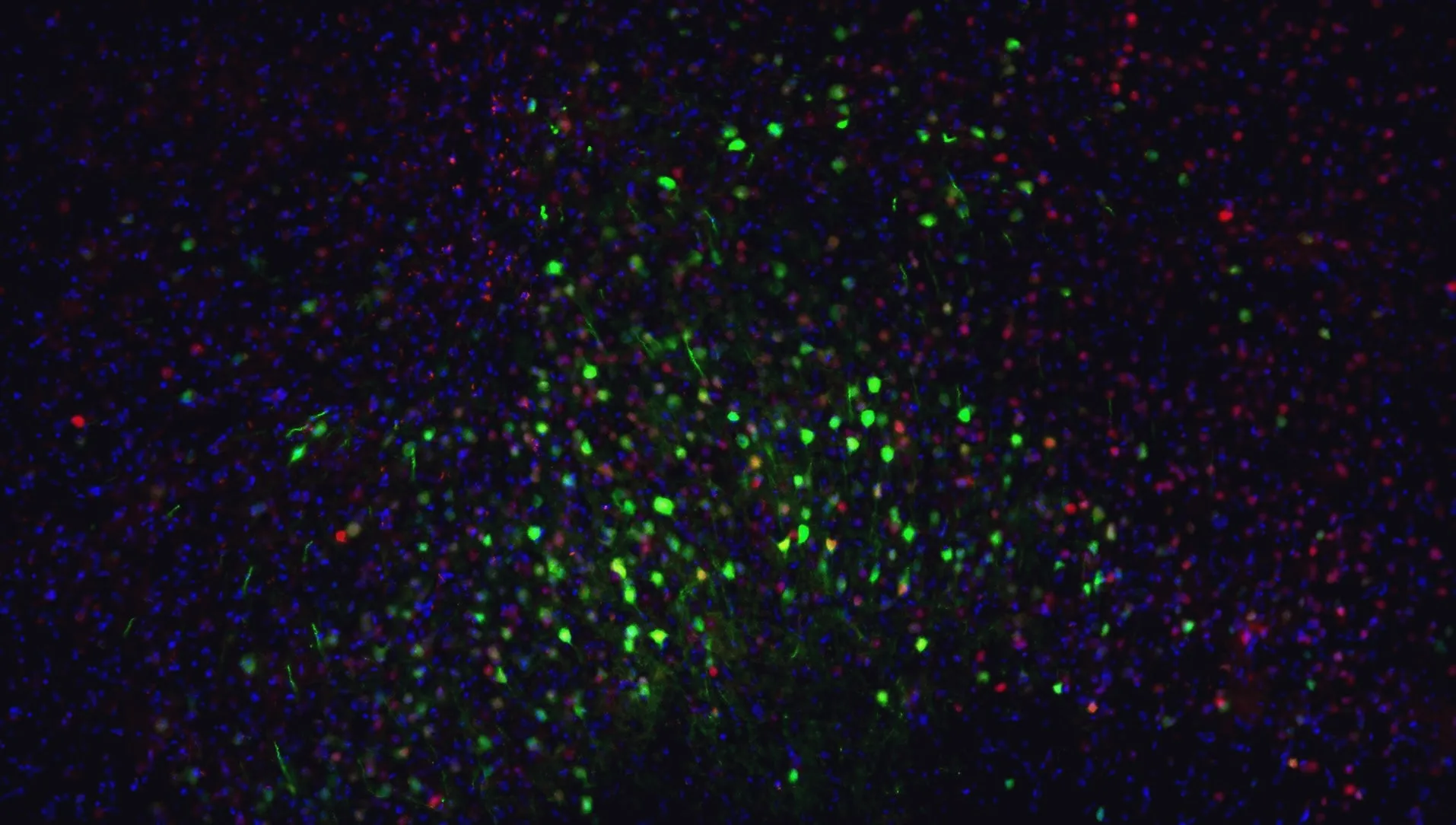
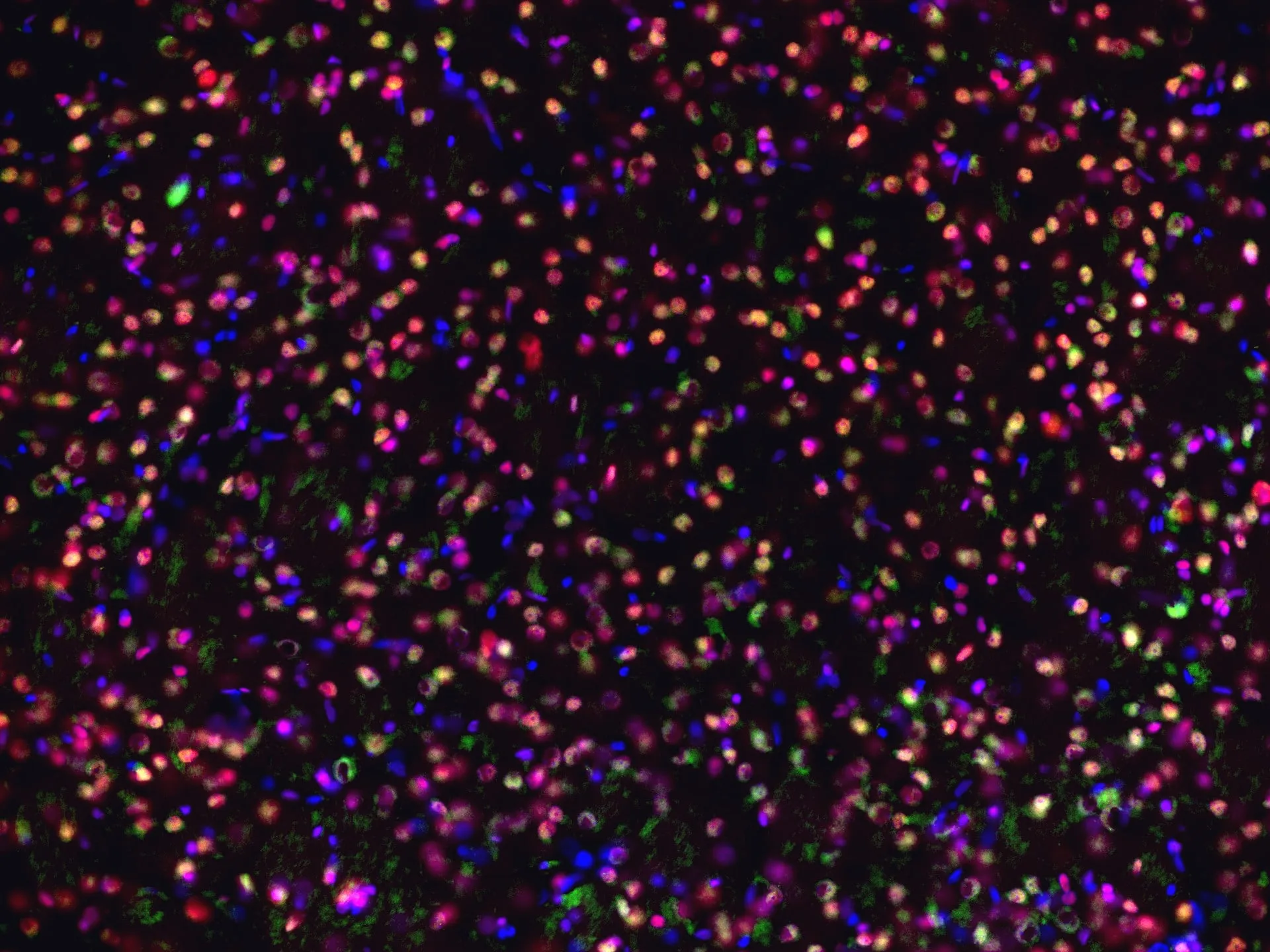
Orbitofrontal cortex, 10×. Imaged in the Wassum Lab.
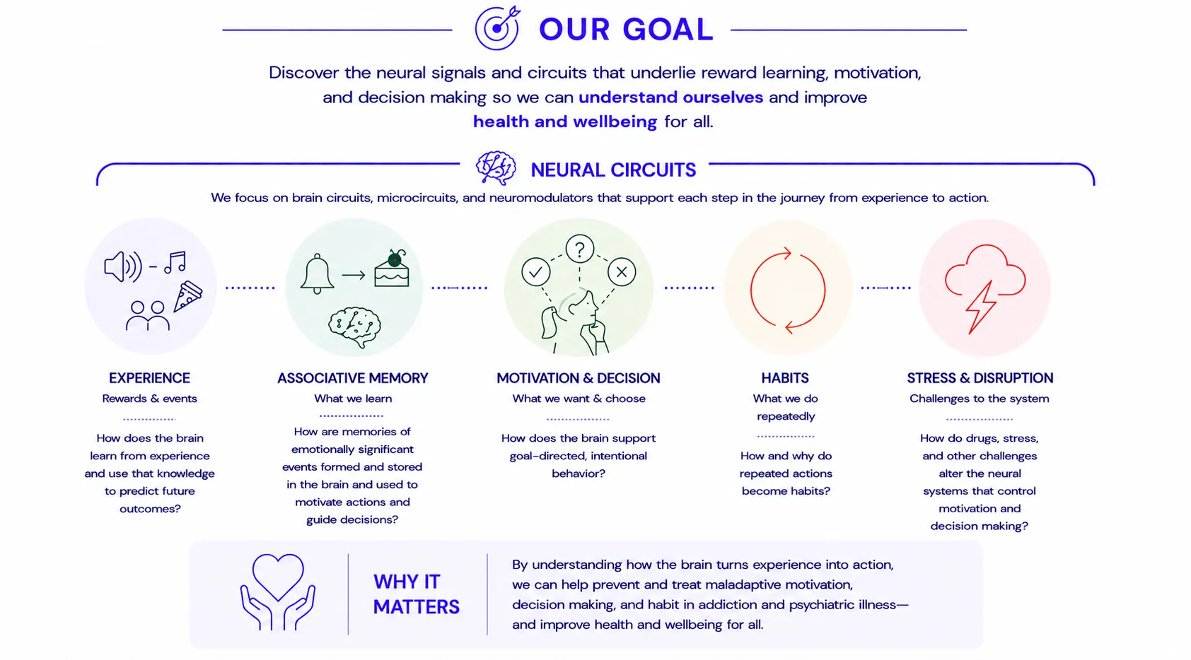
Our goal is to discover the neural signals and circuits that underlie reward learning, motivation, and decision making so that we can capitalize on this information to understand and treat the maladaptive motivation and decision making that characterizes substance use disorder and psychiatric diseases.
We combine ideas and methods across behavioral neuroscience, systems neuroscience, and molecular neuroscience. Our behavioral experiments are grounded in the rich traditions of learning theory and are designed to reveal the computations and psychological processes that shape behavior. We pair these approaches with modern methods for recording, manipulating, and characterizing neural activity to determine how specific signals, cells, and circuits contribute to learning, motivation, and decision making.
We approach science as a collaborative endeavor. We work together to ask rigorous questions, develop as scientists, and make discoveries that matter. We are committed to creating an environment in which people with different backgrounds and perspectives can contribute, learn, and thrive.
Our goals
Discover the neural signals and circuits that underlie reward learning, motivation, and decision making so that we can contribute to the foundation of knowledge that helps us to understand ourselves, why we do the things we do, and how we can harness this information to improve health and wellbeing for all.
Our approach
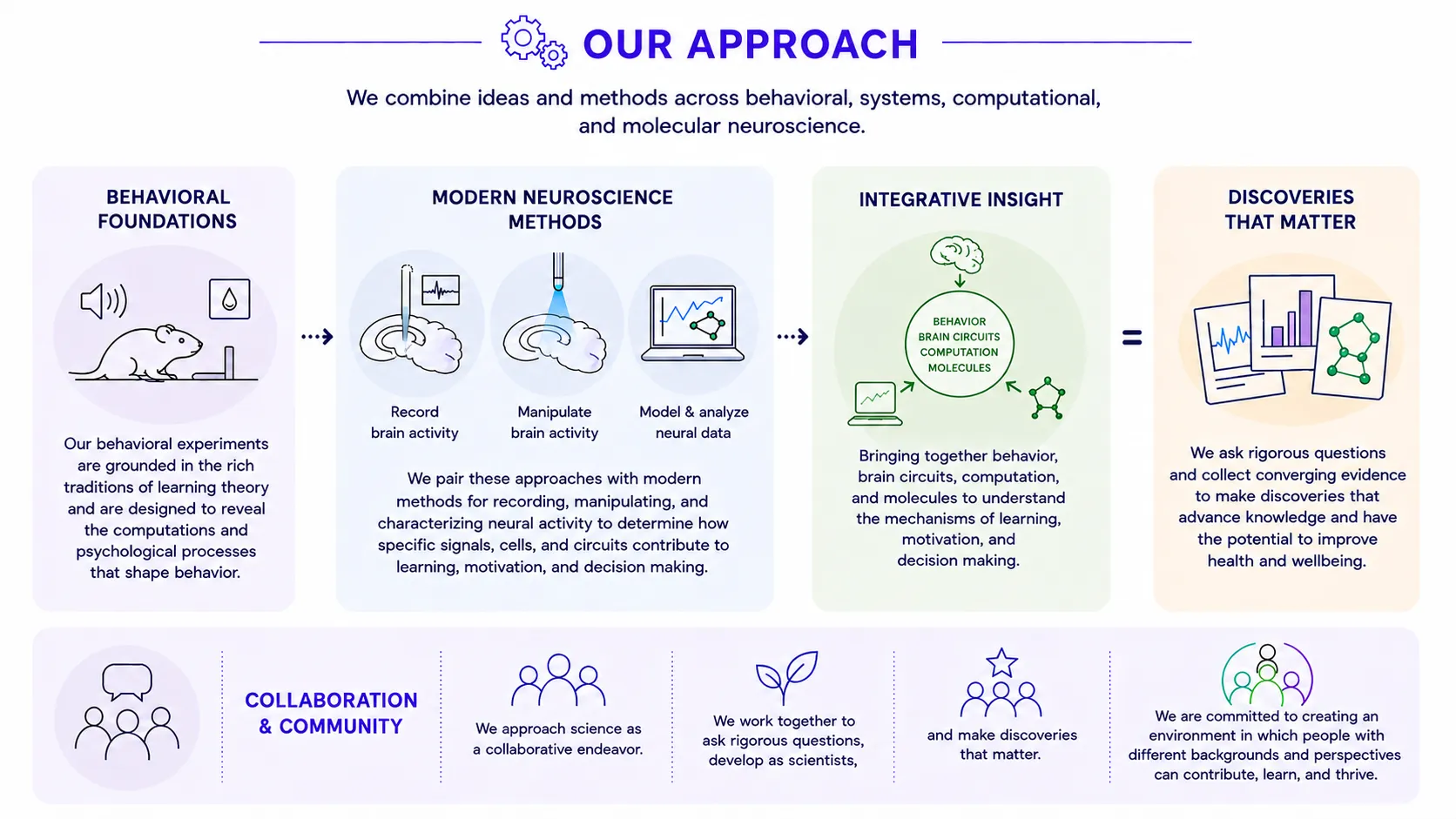
We combine ideas and methods across behavioral, systems, computational, and molecular neuroscience.
Our behavioral experiments are grounded in the rich traditions of learning theory and are designed to reveal the computations and psychological processes that shape behavior. We pair these approaches with modern methods for recording, manipulating, and characterizing neural activity to determine how specific signals, cells, and circuits contribute to learning, motivation, and decision making.
We approach science as a collaborative endeavor. We work together to ask rigorous questions, develop as scientists, and make discoveries that matter. We are committed to creating an environment in which people with different backgrounds and perspectives can contribute, learn, and thrive.
Our current techniques
- Operant, Pavlovian, and other Behavioral assays
- Stress protocols
- Fluorescent biosensors
- Fiber photometry
- Cellular resolution microendoscopic imaging
- Fast-scan cyclic voltammetry
- Viral-mediated gene transfer
- Optogenetics
- Chemogenetics
- Pharmacology
- Permanent and reversible brain lesions
- Circuit tracing and anatomy
- Light and fluorescence microscopy
- Immunohistochemistry
Our funding
NIH 3R01DA035443, R01MH126285, R01DA057084, R01DA058374, R61MH135106 (Suthana PI), T32DA024635